Skip to content

Virgilio Galvis | Ophthalmological Center

REQUEST YOUR APPOINTMENT


[vc_row iw_layout=”wide-bg”][vc_column][vc_row_inner equal_height=”yes” gap=”35″][vc_column_inner width=”1/2″][vc_video link=”https://youtu.be/mgR2yBf0BZ0″][vc_empty_space][/vc_column_inner][vc_column_inner width=”1/2″][inwave_heading preview_style_1=”” title=”{Centro Oftalmológico de Tecnología Avanzada}” align=”left”][vc_column_text]

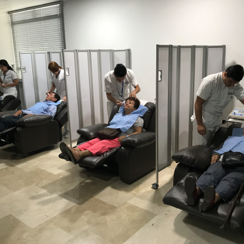
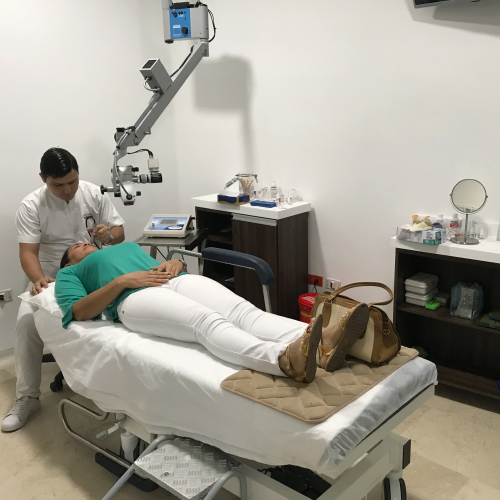
Technology is the most important medical tool in the field of ophthalmology. That's why at the Advanced Technology Ophthalmology Center, called COTA, we have the latest technological equipment for diagnostic tests that help us make an accurate diagnosis for subsequent treatments or surgical and therapeutic procedures.

At COTA, we use state-of-the-art diagnostic tools and are committed to early diagnosis, a vitally important concept for restoring a patient's visual health.

Dentro de los exámenes de apoyo diagnostico que realizamos tenemos los siguientes:[/vc_column_text][vc_btn title=”SOLICITE SU CITA” shape=”round” align=”center” el_class=”popmake-3281 pum-trigger”][/vc_column_inner][/vc_row_inner][vc_row_inner][vc_column_inner][vc_empty_space][vc_column_text]

[slide-anything id=”3305″]

[/vc_column_text][/vc_column_inner][/vc_row_inner][inwave_accordions layout=”accordion2″ preview_style2=””][inwave_accordion_item title=”ABERROMETRY”]Aberrometry allows us to analyze the optical properties of the cornea based on its morphology.

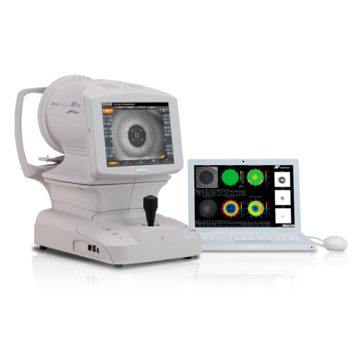
The study of corneal aberrations has multiple uses in ophthalmology: among others, the study of the optical quality of the normal and pathological cornea, the selection of intraocular lenses based on corneal spherical aberration and the application of personalized laser treatments. excimer guided by corneal aberrometry.

The equipment we use is:

Peramis – SCHWIND

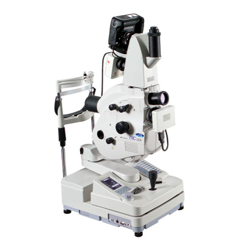
KR1W – TOPCON[/inwave_accordion_item][inwave_accordion_item title=”ANGIOGRAPHY”]Retinal angiography is a diagnostic exam that requires prior pupil dilation and the intravenous administration of a dye used as a contrast medium (Sodium Fluorescein). This will allow the ophthalmologist to evaluate whether the patient has any type of retinal disease and define the course of action to follow.

The equipment we use is:

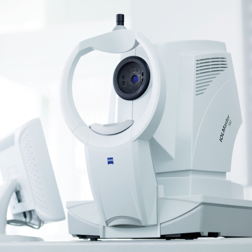
TRC 50 DX – TOPCON[/inwave_accordion_item][inwave_accordion_item title=”OCULAR BIOMETRY”]The ocular biometry examination allows measuring the axial length of the eye, the depth of the anterior chamber and the thickness of the lens

The equipment we use is:

IOL MASTER 700 – ZEISS

LENSTAR L900 – HAAG STREET[/inwave_accordion_item][inwave_accordion_item title=”VISUAL FIELDS – CAMPIMETRY”]The computerized Visual Field exam allows the evaluation of retinal sensitivity, the visual pathway and the optic nerve.

The teams we have are:

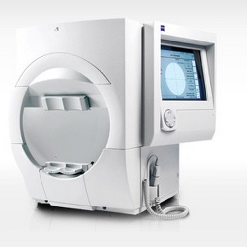
HUMPREY FIEL ANALYSER – ZEISS[/inwave_accordion_item][inwave_accordion_item title=”OCULAR ULTRASOUND”]Ocular ultrasound allows an evaluation of the structures of the posterior segment of the eye, such as the vitreous cavity, retina, choroid, optic nerve and the anterior orbit by means of ultrasound.

The team we have is:

HISCAN-OPTIKON[/inwave_accordion_item][inwave_accordion_item title=”PHOTOS OF THE FUND OF THE EYE – ANTERIOR AND POSTERIOR SEGMENT”]Photographs taken of the most anterior part of the eye are used to document pathologies of the anterior segment and the ocular adnexa.

The teams we have are:

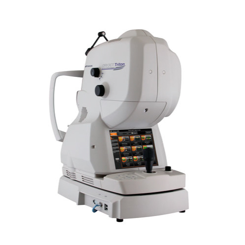
OCT TRITON – TOPCON[/inwave_accordion_item][inwave_accordion_item title=”OPTICAL COHERENCE TOMOGRAPHY”]Optical Coherence Tomography acronym OCT is a non-invasive diagnostic method that allows obtaining high-resolution images of the retina and optic nerve, it does not require contact with the eye as it is based on the reflection of light signals.

The teams we have are:

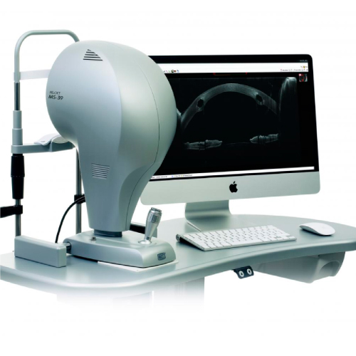
MS39 – CSO

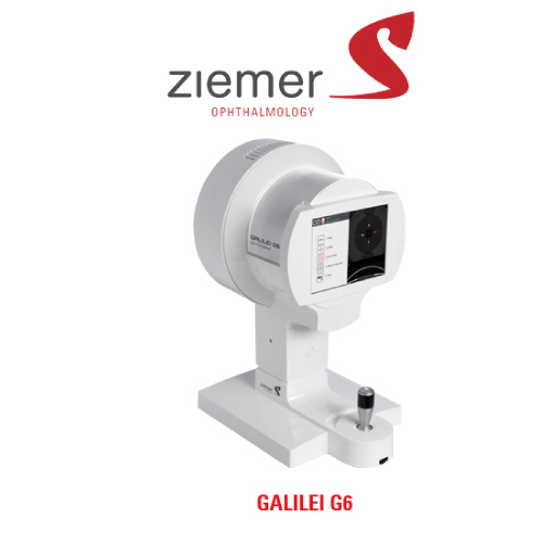
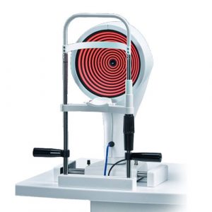
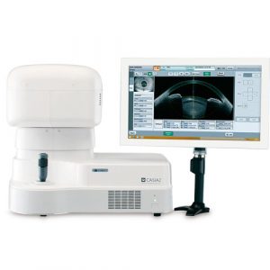
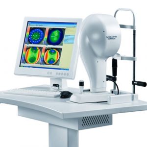
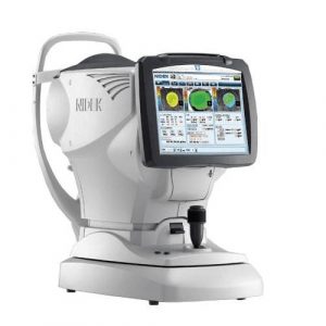
OCT TRITON – TOPCON[/inwave_accordion_item][inwave_accordion_item title=”CORNEAL TOPOGRAPHY”]The Corneal Topography exam performs an analysis of the surface of the cornea, which allows the shape to be evaluated and the power of the different curvatures to be quantified. It also determines whether these are normal deformations or deformations caused by degenerative diseases such as keratoconus.

The teams we have are:

SIRIUS – SCHWIND

MS39 – CSO

GALILEI G6 – ZIEMER

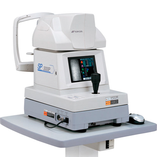
OPD SCAN III – NIDEK [/inwave_accordion_item][inwave_accordion_item title=”ENDOTHELIAL CELL COUNT”]The endothelial cell count test allows the evaluation of the quantity and quality of the cells of the corneal endothelium or inner layer of the cornea. It is necessary to assess the health of the cornea, the presence of corneal dystrophies, the possibility of rejection in a corneal transplant, and whether or not the cornea is resistant to certain procedures.

The equipment we have is:

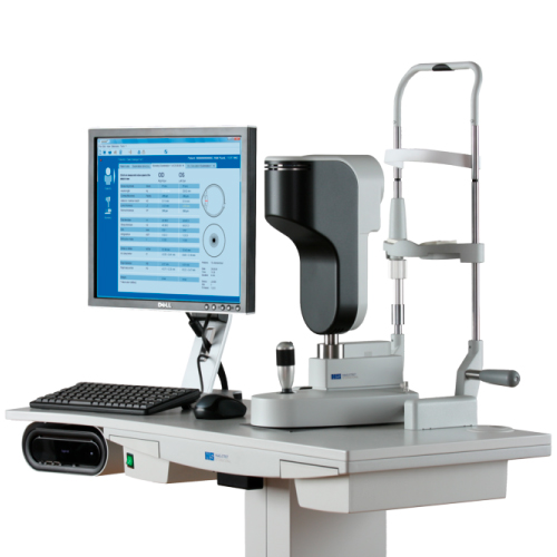
SP3000 – TOPCON[/inwave_accordion_item][inwave_accordion_item title=”UBM ULTRABIOMICROSCOPY ULTRASOUND”]UBM Ultrasound allows for the evaluation of the structures of the anterior segment of the eye through high-resolution images obtained by ultrasound.

The equipment we have is:

HISCAN – OPTIKON[/inwave_accordion_item][inwave_accordion_item title=”LASER PHOTOCOAGULATION”]Laser photocoagulation is an eye surgery that uses a laser to shrink or destroy abnormal structures in the retina, or to intentionally cause scarring.

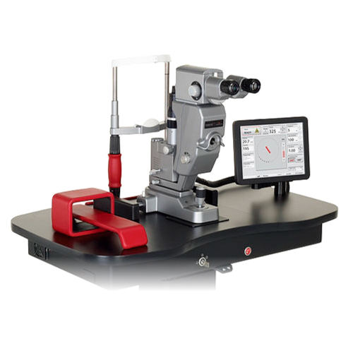
The equipment we have is: LASER PASCAL STREAM LINE 577 – TOPCON[/inwave_accordion_item][inwave_accordion_item title=”IRIDOTOMY AND CAPSULOTOMY”]Iridotomy is a procedure performed with a laser to facilitate the drainage of the aqueous humor. It is a treatment performed on eyes with a narrow angle (occludable angle) for preventive purposes: to avoid an episode of glaucoma, that is, a sudden rise in intraocular pressure due to pupillary block.

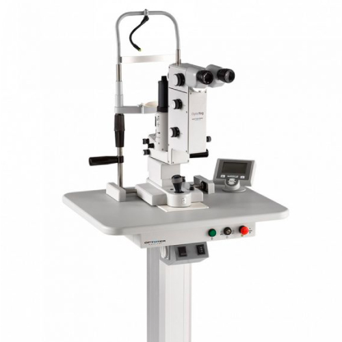
Capsulotomy is the technique that eliminates the opacity of the capsule surrounding an implanted intraocular lens; this opacity is caused by the proliferation of residual cells.

The equipment we have is: YAG LASER – CSO[/inwave_accordion_item][inwave_accordion_item title=”OCULAR ELECTROPHYSIOLOGY”]It is the study of the electrical potentials of the cells and tissues of the retina.
Electrophysiological examination consists of evaluating the function of the visual pathway from the photoreceptors (rods and cones) of the retina to the visual cortex of the brain.

Basic tests in electrophysiology: Electroretinogram, Visual Evoked Potential, Electrooculogram.


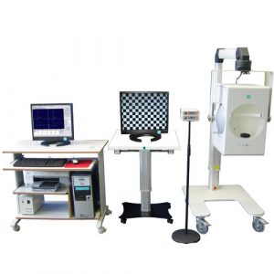
El equipo que tenemos es: RETI-PORT/SCAN21 Gamma Plus2[/inwave_accordion_item][/inwave_accordions][vc_empty_space][vc_btn title=”SOLICITE SU CITA” shape=”round” align=”center” el_class=”popmake-3281 pum-trigger”][/vc_column][/vc_row]