TRC 50 DX RETINAL CAMERA
Ultrasound
Ultrasonic Biomicroscopy
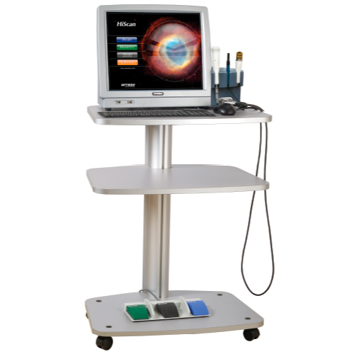
The high technological content of HiScan Touch, A/B-Scan ultrasound and UBM, allows exceptional performance in the diagnosis and measurement of the anterior and posterior sections thanks to the extreme quality of the images, guaranteed by the use of advanced technology. It is a stand-alone system that uses advanced software and has a full range of probes. Thanks to its extremely high sensitivity, the 12 MHz B-Scan probe allows the acquisition of detailed images of the posterior segment and especially the vitreous and orbit. HiScan Touch is the only ultrasound with an integrated system for 3D reconstruction of the anterior and posterior segment.
It is a diagnostic imaging technique that studies the structures of the eyeball and the structures attached to the orbit (muscles, lacrimal gland, orbital fat, etc.) using ultrasound. Like abdominal ultrasounds or other parts of the body, to perform an ocular ultrasound, a gel is spread on the patient's skin (in this case the eyelid, although it can be performed directly on the eyeball) and the ultrasound probe with said gel to obtain the images.
Ultrasonic Biomicroscopy (UBM) is a high-resolution ultrasound technique that allows the structures of the anterior segment of the eye to be analyzed in detail, structures that are otherwise hidden because they cannot be seen by other means or equipment. Although it is a slow method to perform compared to other techniques for studying the anterior pole, it allows detailed analysis of the posterior ocular chamber. It is considered a very useful technique in the implantation of phakic intraocular lenses and is essential in the study of certain tumors of the anterior ocular segment. Other important utilities are glaucoma cases and their postoperative follow-up. It is a useful technique, likewise, in patients who present media opacity and therefore the exploration of the anterior ocular segment by other means is difficult.
