
TRC 50 DX RETINAL CAMERA
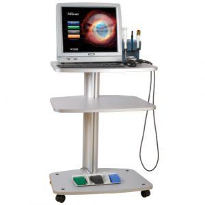
- Ultrasound
- Ultrasonic Biomicroscopy
It is a diagnostic imaging technique that studies the structures of the eyeball and the structures adjacent to the orbit (muscles, tear gland, orbital fat, etc.) using ultrasound. Similar to ultrasounds of the abdomen or other parts of the body, an ocular ultrasound involves applying a gel to the patient's skin (in this case, the eyelid, although it can be performed directly on the eyeball) and placing the ultrasound probe in contact with the gel to obtain images.
Ultrasound biomicroscopy (UBM) is a high-resolution ultrasound technique that allows for detailed analysis of the anterior segment of the eye, structures that are otherwise hidden because they cannot be seen by other means or equipment. Although it is a slow method compared to other techniques for studying the anterior pole, it allows for detailed analysis of the posterior chamber of the eye. It is considered a very useful technique in the implantation of phakic intraocular lenses and is essential in the study of certain anterior segment tumors. Other important uses include cases of glaucoma and their postoperative follow-up. It is also a useful technique in patients with opacity of the media, making examination of the anterior segment of the eye difficult by other means.
Aberrometry allows us to analyze the optical properties of the cornea based on its morphology. The study of corneal aberrations has multiple uses in ophthalmology: among others, the study of the optical quality of normal and pathological cornea, the selection of intraocular lenses based on corneal spherical aberration, and the application of personalized excimer laser treatments guided by corneal aberrometry. The aberrometric map can be used to design a customized ablation map for use on an excimer laser platform to correct the patient's higher-order aberrations and improve visual quality after corneal refractive surgery.