[vc_row iw_layout=”wide-bg”][vc_column][vc_row_inner equal_height=”yes” gap=”35″][vc_column_inner width=”1/4″][vc_wp_custommenu title=”ENFERMEDADES OCULARES” nav_menu=”41″][/vc_column_inner][vc_column_inner width=”3/4″][inwave_heading preview_style_1=”” title=”{RETINA Y VÍTREO}”][vc_column_text]La especialidad denominada retina y vítreo, trabaja en la retina que es un tejido sensible a la luz situado en la superficie interior del ojo, es como una pantalla donde se proyectan las imágenes.
Light striking the retina triggers chemical and electrical reactions that are converted into nerve impulses that travel to the brain through the optic nerve. The retina has a very complex structure, composed of several layers of neurons interconnected by synapses.
The retina is the neural tissue that covers the inner surface of the eye, as if it were a room and the retina were the wallpaper.
The retina is not a normal tissue, but a small part of the brain responsible for collecting images. Because it's composed of brain cells organized into highly structured layers, anything that affects it can cause serious visual problems.
Some retinal and vitreous diseases:
[/vc_column_text][/vc_column_inner][/vc_row_inner][vc_row_inner equal_height=”yes” gap=”35″][vc_column_inner width=”1/3″][vc_empty_space][/vc_column_inner][vc_column_inner width=”1/3″][inwave_heading preview_style_1=”” title=”{Enfermedades de la Retina}”][/vc_column_inner][vc_column_inner width=”1/3″][/vc_column_inner][vc_column_inner][/vc_column_inner][/vc_row_inner][vc_row_inner][vc_column_inner width=”1/2″][inwave_item_info style=”style1_4″ preview_style4=”” title=”{Retinopatía Diabética}” description=”Daño producido a causa de la descompensación metabólica de la diabetes.” icon_size=”50″ align=”center” img=”3323″][/vc_column_inner][vc_column_inner width=”1/2″][inwave_item_info style=”style1_4″ preview_style4=”” title=”{Agujero Macular}” description=”Pequeña ruptura en la mácula, la parte del ojo responsable de la visión central.” icon_size=”50″ align=”center” img=”3320″][/vc_column_inner][/vc_row_inner][vc_row_inner][vc_column_inner width=”1/2″][inwave_item_info style=”style1_4″ preview_style4=”” title=”{Desprendimiento y Desgarro de Retina}” description=”Separación espontánea de la retina, puede causar ceguera.” icon_size=”50″ align=”center” img=”3319″][/vc_column_inner][vc_column_inner width=”1/2″][inwave_item_info style=”style1_4″ preview_style4=”” title=”{Edema Macular}” description=”Acumulación en la mácula de líquido en la retina.” icon_size=”50″ align=”center” img=”3322″][/vc_column_inner][/vc_row_inner][vc_row_inner][vc_column_inner width=”1/2″][inwave_item_info style=”style1_4″ preview_style4=”” title=”{Membranas epiterrianas}” description=”Desarrollo de una membrana celular translúcida sobre la zona central de la retina.” icon_size=”50″ align=”center” img=”3319″][/vc_column_inner][vc_column_inner width=”1/2″][inwave_item_info style=”style1_4″ preview_style4=”” title=”{Complicaciones por la uveítis}” description=”Factores de riesgo por padecer de uveitis.” icon_size=”50″ align=”center” img=”3322″][/vc_column_inner][/vc_row_inner][vc_empty_space][vc_row_inner gap=”35″][vc_column_inner width=”1/2″][vc_video link=”https://www.youtube.com/watch?v=Q63e3knahyE”][/vc_column_inner][vc_column_inner width=”1/2″][inwave_heading preview_style_1=”” title=”{RETINOPATÍA DIABÉTICA}” sub_title=”Es la enfermedad vascular más frecuente de la retina, Se origina por el daño producido en los vasos retinianos a causa de la descompensación metabólica de la diabetes. Con lleva a una pérdida de visión que, en ocasiones, puede llevar a la ceguera. Es la principal causa de ceguera en el mundo.”][inwave_accordions layout=”accordion2″ preview_style2=”” item_active=”-1″][inwave_accordion_item title=”Síntomas”]Con el mantenimiento de elevados niveles de glicemia, las paredes de los vasos retinianos se alteran y se vuelven más permeables, dejando pasar fluido al espacio extracelular.
In more advanced cases, there is a proliferation of abnormal blood vessels that cause bleeding.
The presence of blood in the vitreous space (a transparent gel that fills the eyeball) causes it to become opaque, causing a decrease in vision, which generally occurs suddenly.
The more years of evolution there is, the greater the chances of developing retinopathy.
Often, the patient is unaware of the disease until the damage is severe, hence the importance of regular retinal examinations.
Symptoms of diabetic retinopathy may include:
- Blurred vision and gradual loss of vision
- Vision of spots or “floaters”
- Shadows or lost areas of vision
- Difficulty seeing at night
[/inwave_accordion_item][inwave_accordion_item title=”Treatment”]The diabetic population must strictly control their blood sugar, blood pressure, cholesterol and triglycerides.
Other factors that negatively influence diabetic retinopathy include obesity, smoking, and a sedentary lifestyle.
These patients require periodic retinal examinations, since diabetic retinopathy generally does not cause symptoms until the injury is severe.
Retinopathy can affect the macula (central area of the retina responsible for detailed vision) or its periphery.
Depending on the affected area and the degree of development of the disease, specialists have different treatment options, such as laser photocoagulation, intravitreal injections or surgery (vitrectomy).
Otras complicaciones visuales asociadas a la diabetes, como el glaucoma o las cataratas, requieren de tratamientos específicos.[/inwave_accordion_item][/inwave_accordions][/vc_column_inner][/vc_row_inner][vc_row_inner gap=”35″][vc_column_inner width=”1/2″][inwave_heading preview_style_1=”” title=”{AGUJERO MACULAR}” sub_title=”El agujero macular es por lo general una consecuencia del envejecimiento, por lo que suele afectar a personas mayores de 60 años. El proceso de envejecimiento tiene dos efectos concretos en la estructura del ojo. Por un lado, la mácula, que es la parte central de la retina, se hace cada vez más delgada. Por otro, se contrae el humor vítreo, una sustancia gelatinosa que en condiciones normales ocupa todo el interior del globo ocular. A lo largo de este proceso cabe la posibilidad de que una parte del humor vítreo permanezca adherido a la mácula en vez de despegarse, de modo que tira de ella. Esto, unido al adelgazamiento de la mácula, favorece que la tensión generada produzca el desgarro de la parte central de la retina. Un agujero macular es una pequeña ruptura en la mácula, la parte del ojo responsable de la visión central. Además de la edad, la miopía, el trauma ocular, ciertas lesiones oculares o un proceso inflamatorio ocular de larga duración son otros factores de riesgo de sufrir un agujero macular.”][inwave_accordions layout=”accordion2″ preview_style2=”” item_active=”-1″][inwave_accordion_item title=”Síntomas”]En la fase inicial de formación del agujero macular se produce una alteración de la visión que se traduce en visión borrosa, neblinosa o con ondulaciones. Si éste se agranda aún más puede aparecer un punto negro y distorsión en la visión central, que será tanto mayor cuanto mayor sea el área de la mácula que ha resultado afectada.
Any of these symptoms should lead you to immediately consult an ophthalmologist for an in-depth study of the structure of the eye and if the diagnosis is confirmed, immediate treatment can be carried out. [/inwave_accordion_item] [inwave_accordion_item title = “Treatment”] Treatment of macular holes is always surgical and is performed through a procedure called vitrectomy, in order to detach the vitreous humor adhered to the macula and depending on the size of the hole, different surgical techniques will be performed to try not only to close it, but also to achieve the regeneration of the layers of the retina.
[/inwave_accordion_item][/inwave_accordions][/vc_column_inner][vc_column_inner width=”1/2″][vc_video link=”https://www.youtube.com/watch?v=COu1kB-5Cg0″][/vc_column_inner][/vc_row_inner][vc_row_inner gap=”35″][vc_column_inner width=”1/2″][vc_video link=”https://www.youtube.com/watch?v=EEAoGa6zNpQ”][/vc_column_inner][vc_column_inner width=”1/2″][inwave_heading preview_style_1=”” title=”{DESPRENDIMIENTO DE RETINA}” sub_title=”El desprendimiento de retina es una enfermedad ocular que se produce por la separación espontánea de la retina neurosensorial del epitelio pigmentario. Al no causar dolor y, en muchos casos, no ir acompañado al inicio de pérdida de visión, es importante estar alerta ante los síntomas de un desprendimiento de retina, aunque éstos sean aparentemente inofensivos.”][inwave_accordions layout=”accordion2″ preview_style2=”” item_active=”-1″][inwave_accordion_item title=”Síntomas”]Estos síntomas, que suelen aparecer sucesivamente, son:
- Floaters (black spots that move when you move your eye). These are caused by changes in the vitreous.
- Flashing light. This is a more serious symptom, reflecting the existence of traction on the retina. It usually appears after a tear has already occurred.
- Vision of a black curtain falling across an area of the visual field. This occurs when retinal detachment is already present, so immediate consultation with an ophthalmologist is necessary.
- Image distortion and subsequent significant loss of visual acuity. This symptom occurs if the central area of the retina (macula) is damaged.
It is important for the at-risk population to undergo regular eye exams, at least once a year.
Furthermore, the sudden appearance of floaters or a sudden increase in existing floaters, as well as the appearance of flashes of light or any other of the symptoms described, should be reason for urgent consultation with an ophthalmologist.[/inwave_accordion_item][inwave_accordion_item title=”Treatment”]It is very important to make a diagnosis as quickly as possible, since the chances of improvement are greater if the macula or central area of the retina does not become detached.
Preventive laser treatment is advisable when retinal tears are present, even if they have not yet caused a detachment.
This preventative laser treatment can also be useful for high-risk patients with peripheral retinal degenerative lesions that could lead to retinal tears.
There are different surgical techniques, depending on the degree and stage of retinal detachment:
- Laser photocoagulation. Lasers cause controlled burns around the detached area. These burns eventually heal and seal the retinal tear, preventing vitreous humor from infiltrating between the two layers.
- Vitrectomy. This involves removing the vitreous humor from inside the eye. The retina is then applied using heavy liquids and laser treatment from inside the eye.
- Scleral surgery. A solid silicone band is placed around the outermost layer of the eye wall (the sclera) to maintain external pressure on the eyeball, which facilitates closure of the tear.
[/inwave_accordion_item][/inwave_accordions][/vc_column_inner][/vc_row_inner][vc_row_inner gap=”35″][vc_column_inner width=”1/2″][inwave_heading preview_style_1=”” title=”{EDEMA MACULAR}” sub_title=”El edema macular se produce por la acumulación en la mácula de líquido que sale de los vasos sanguíneos que alimentan la retina, bien porque éstos son anómalos o porque sus paredes son demasiado delgadas, lo que causa su dilatación y aumento de la permeabilidad. Lo habitual es que se trate de una complicación de diferentes patologías oculares, como la retinopatía diabética, trombosis venosa de la retina, degeneración macular asociada a la edad, uveítis, entre otras. Las causas más frecuentes son la retinopatía diabética y el edema secundario a trombosis venosas de la retina.”][inwave_accordions layout=”accordion2″ preview_style2=”” item_active=”-1″][inwave_accordion_item title=”Síntomas”]Hay que tener en cuenta que la mácula es la parte central de la retina y permite enfocar y ver con detalle cualquier objeto, permitiendo realizar actividades como leer, ver la cara de las personas, coser, enhebrar una aguja, etc.
Consequently, the symptoms of macular edema affect central vision, which may include blurred vision, line distortion, and altered shape of objects. If not treated properly, the patient may develop severely limited central vision. [/inwave_accordion_item] [inwave_accordion_item title=”Treatment”] Treatment for macular edema will depend on the cause, the degree of progression, and whether it is focal or diffuse; it will be determined based on the characteristics of each patient. Therapeutic options include pharmacological treatment with eye drops, or intraocular and periocular injections. These medications include antiangiogenic and steroid agents, which act on endothelial growth factor receptors.
Intraocular drugs can control inflammation in the center of the macula.
Other treatment options focus on laser photocoagulation of the blood vessel sites where fluid loss occurs, which accumulates in the macula, impairing its function.
In the most severe cases, vitrectomy surgery may be necessary.
La vitrectomía es una intervención quirúrgica en la que se extrae el gel transparente que llena el globo ocular (humor vítreo) para acceder a la retina y tratar los vasos anómalos y eliminar las posibles bandas fibrosas que puedan ejercer una tracción sobre la retina, con el subsiguiente riesgo de que se desgarre y se produzca un desprendimiento de retina.[/inwave_accordion_item][/inwave_accordions][/vc_column_inner][vc_column_inner width=”1/2″][vc_video link=”https://www.youtube.com/watch?v=JPsejooQ_QQ”][/vc_column_inner][/vc_row_inner][vc_row_inner gap=”35″][vc_column_inner width=”1/2″][vc_video link=”https://youtu.be/k0x1XzYFkLk”][/vc_column_inner][vc_column_inner width=”1/2″][inwave_heading preview_style_1=”” title=”{EDEMA MACULAR DIABÉTICO}” sub_title=”El edema macular ocurre en personas que tienen vasos sanguíneos con fugas en la retina. Esto se conoce como retinopatía diabética. El edema macular ocurre cuando estos vasos sanguíneos dañados pierden líquido en la mácula, lo que hace que se hinche. El edema macular puede causar visión borrosa y desteñida.”][inwave_accordions layout=”accordion2″ preview_style2=”” item_active=”-1″][inwave_accordion_item title=”Sintomas”]En sus primeras etapas, el edema macular a menudo no muestra signos de advertencia. Si no se trata, el edema macular puede empeorar, causando cambios en su visión y posiblemente conducir a la pérdida de la visión. La detección temprana del edema macular es la mejor manera de prevenir la pérdida de visión.
Here's what to look for:
- Small patches of vision loss
- The colors appear to be “washed out” or changed
- Straight lines look bent or crooked
Even if you don't have symptoms, you should have a dilated eye exam every year. A dilated eye exam is the only way to know if diabetes is affecting your retina or macula.
RISK FACTORS
The following list includes some of the factors that may put you at greater risk for macular edema:
- Having diabetes for more than 10 years.
- High blood sugar levels and high fasting blood glucose levels.
- Other risk factors for macular edema include high blood pressure, high cholesterol, and smoking.
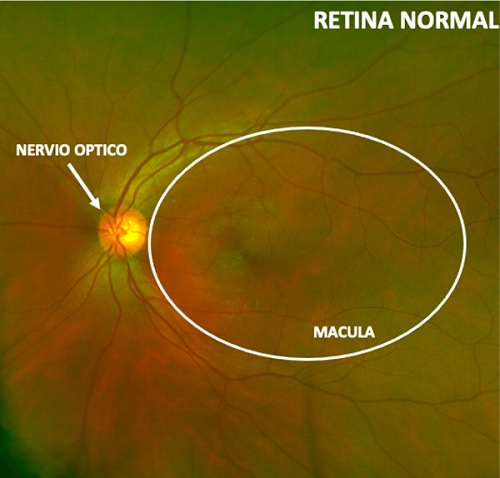
[/inwave_accordion_item][inwave_accordion_item title=”What does a healthy retina look like?”]In a healthy retina, all the structures are visible without any alteration.
A healthy macula looks like a gap in the retina.
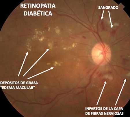
 [/inwave_accordion_item][inwave_accordion_item title=”What does a retina with macular edema look like?”]In an eye with macular edema, blood vessels leak. Fluid builds up in the retina and macula. This causes the macula to swell and bulge. With a swollen macula, it can be difficult to see fine details.
 [/inwave_accordion_item][/inwave_accordions][/vc_column_inner][/vc_row_inner][vc_row_inner gap=”35″][vc_column_inner width=”1/2″][inwave_heading preview_style_1=”” title=”{UVEITIS}” sub_title=”La uveítis es una inflamación de la úvea, membrana que envuelve el interior del globo ocular y que se encarga de la nutrición del ojo por dentro. La uveítis es una de las causas de ceguera más importantes en el mundo, debido al contacto íntimo que tiene la úvea con partes importante del ojo como el nervio óptico y la retina. Lo mas importante de las uveítis, es que en un gran porcentaje se encuentran asociadas a enfermedades sistémicas que aun no se han manifestado y/o diagnosticado.”][inwave_accordions layout=”accordion2″ preview_style2=”” item_active=”-1″][inwave_accordion_item title=”Sintomas”]Los síntomas de la uveítis son diferentes según la zona de la úvea que se encuentra afectada.
- If it is the anterior part, we can notice increased sensitivity to light (photophobia), redness of the eyes, blurred vision or eye pain.
- If the affected area is the back, we may not notice pain, although we may experience loss of vision or seeing floaters.
There are several types of factors that cause uveitis:
- Infectious diseases such as toxoplasmosis (the most common cause of uveitis in Colombia). This process results in the gradual destruction of the retina, which, if it affects the macula (central part of the retina), can cause significant, irreversible vision loss.
- Rheumatic diseases of inflammatory, non-degenerative origin, which primarily affect young people, such as rheumatoid arthritis, ankylosing spondylitis, among others.
- Autoimmune diseases caused by viruses, germs, or environmental factors, such as sarcoidosis or Behçet's disease, a disease of unknown origin that causes sores and skin changes.
- Traumas
Most people affected by uveitis are between 20 and 50 years old.[/inwave_accordion_item][inwave_accordion_item title=”Treatment”]There are different treatments depending on the type and location of the uveitis:
- Anterior uveitis is treated, in most cases, with anti-inflammatory eye drops.
- Non-infectious posterior uveitis is treated with steroids administered orally or through injections around the eye.
- Chronic forms of uveitis may require the use of immunomodulatory drugs.
- Surgery is not a common treatment to cure uveitis, but it can be effective in controlling associated complications that generated uveitis that was not diagnosed and/or treated in time, such as cataracts or glaucoma (affecting the anterior segment), retinal detachment, vitreous opacity or macular edema (affecting the fundus of the eye).
Habitualmente es necesaria la colaboración de un médico internista, infectólogo y/o reumatólogo que complemente el estudio y tratamiento de la enfermedad causante de la uveítis. En algunos casos, el tratamiento de estas patologías o infecciones puede ayudar a prevenirla.[/inwave_accordion_item][/inwave_accordions][/vc_column_inner][vc_column_inner width=”1/2″][vc_video link=”https://youtu.be/diNfpfHz2Hc”][/vc_column_inner][/vc_row_inner][vc_row_inner][vc_column_inner][vc_btn title=”SOLICITE SU CITA” shape=”round” align=”center” el_class=”popmake-3281 pum-trigger”][/vc_column_inner][/vc_row_inner][/vc_column][/vc_row]