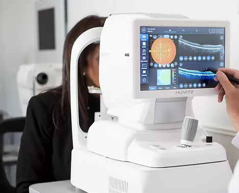
MS-39
Anterior Segment Optical Coherence Tomography
It is the most advanced device for anterior segment analysis. MS-39 combines Placido disk corneal topography with high-resolution OCT-based anterior segment tomography. The clarity of the 16 mm cross-sectional images, along with the many details of the corneal structure and layers revealed by MS-39, will be appreciated by anterior segment specialists. MS-39 provides information on pachymetry, elevation, curvature, and dioptric power of both corneal surfaces. In addition to clinical anterior segment diagnostics, MS-39 can be used in corneal surgery for refractive surgery planning. An IOL calculation module, based on ray tracing techniques, is also available. Additional tools allow MS-39 to perform precise pupil diameter measurements and advanced tear film analysis.
Optical coherence tomography (OCT) allows us to obtain high-resolution cross-sectional images of the anterior and posterior segments of the eye noninvasively using optical interferometry. It has now become a very useful tool for the ultrastructural study of ocular anatomy. Anterior segment OCT is used in the follow-up of patients undergoing refractive surgery, intrastromal rings, corneal cross-linking, corneal transplants, phakic intraocular lenses, and in patients undergoing filtering surgery for glaucoma. In the field of cataract surgery, OCT allows us to accurately analyze the architecture of incisions, as well as the relationships between the intraocular lens and the posterior capsule. Anterior segment OCT is very useful in the analysis and evaluation of anterior segment tumors and cysts, conjunctival tumors, and various corneal conditions such as dystrophies, degenerations, and infections.