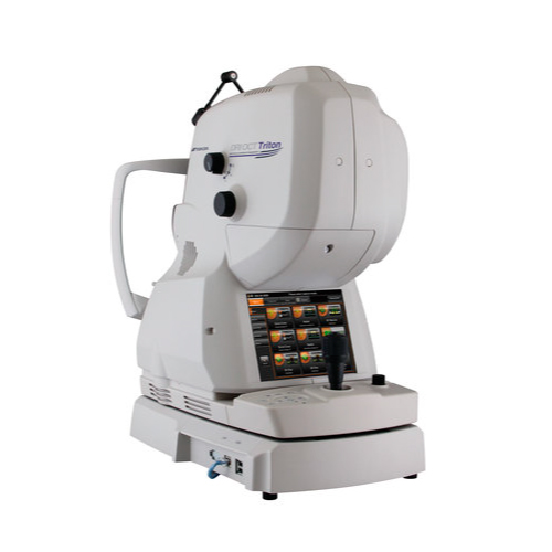

OCT TRITON
- Optical Coherence Tomography – Posterior Segment
- Optical Coherence Tomography – Angiography
TOPCON is the world’s first to introduce a combined anterior and posterior segment Swept Source OCT, the DRI OCT Triton. The DRI OCT Triton features high-resolution color retinography and FA and FAF imaging. FA and FAF images are standard. Swept Source Technology and 1050nm Wavelength The Swept Source OCT offers a significant improvement over conventional OCT. Thanks to the long-wavelength scanning light (1050nm), penetration into the deeper layers of the eye is optimized. It also penetrates better through cataracts, hemorrhages, blood vessels, and the sclera.
Optical Coherence Tomography, or OCT, is a noninvasive imaging technique that allows for detailed examination of the retina, specifically the macula and optic nerve. It is highly useful for the early detection of macular pathologies such as ocular diabetes or age-related macular degeneration, as well as for more effective treatment. OCT provides a wealth of information, allowing for the identification of the stage of the disease and the changes occurring between visits. In the field of the retina, OCT allows for the detection of the most common pathologies such as macular edema, macular holes, epiretinal membranes, retinal dystrophies, retinal vein occlusions, among others.
Optical coherence tomography angiography is a new imaging method based on high-resolution images that demonstrate retinal and choroidal circulation without contrast. This reliable, real-time, and noninvasive method allows visualization of non-irrigated areas in the macular region associated with diabetic retinopathy.